
Category: Pet Health

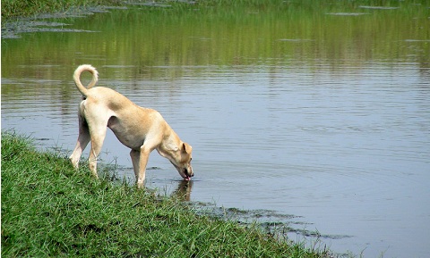
Acute Caudal Myopathy (Limber Tail)
My 10-year-old Labrador retriever suddenly stopped wagging his tail. It was really droopy, and my veterinarian says he has limber tail. What is that?
The term limber tail is one of several slang terms that apply to a condition that is technically called acute caudal myopathy. Some of the other terms you might hear that apply to this include:
- swimmer’s tail
- cold water tail
- dead tail
- broken tail
- limp tail
- rudder tail
- broken wag
Working dogs and active hunting dogs seem to be at greatest risk for developing this condition, but it can happen to any breed.
Is this a true medical condition?
Yes. Acute caudal myopathy typically results from overuse of the tail, causing a strain or sprain of the muscle groups used for tail wagging. Possible scenarios leading to limber tail include hard/vigorous play within the previous 24 hours, prolonged swimming, or active hunting within the past few days. Your dog may act fine immediately following activity but will wake up in pain the next day. The key risk factors appear to be overexertion and/or exposure to very cold water or cold weather.
How is limber tail diagnosed?
Typically, limber tail is diagnosed by connecting the dots between your dog’s symptoms and recent high activity, in addition to a careful evaluation of your dog’s tail by your veterinarian.
Your dog may have difficulty rising because dogs use their tails for balance. Likewise, your dog may have difficulty finding a comfortable position in which to sit and you may see him shifting his weight from side to side. The tail may droop limply between your dog’s rear legs, or it may stick straight out behind him for a short distance before drooping. Your dog may be so distracted by his pain that he might not eat and may be reluctant to squat to defecate.
The veterinary examination will include a careful palpation of the tail starting at the base (up by the pelvis) and proceeding down the entire length. The goal is to locate the discomfort and rule out any other problems that might explain the symptoms.
What else can explain these symptoms?
Other medical problems that resemble limber tail include:
- tail fracture
- lower back pain from a diseased intervertebral disk or osteoarthritis
- infection or inflammation of the anal glands
- prostate disease
The fact that other medical problems can look similar to limber tail reinforces the need for a thorough examination by your veterinarian.
How is limber tail treated?
Uncomplicated acute caudal myopathy is treated with rest and anti-inflammatory medication (e.g., meloxicam, brand name Metacam®). Please only use medication that has been prescribed by your veterinarian. Most dogs are back to normal within a few days to a week. Just because your dog developed limber tail once does not mean that it will happen again when he returns to his favorite activities. You do not need to prevent your dog from doing the things that feed his soul!
Contributors: Tammy Hunter, DVM; Robin Downing, DVM, CVPP, CCRP, DAAPM
© Copyright 2019 LifeLearn Inc. Used and/or modified with permission under license.

Eye Discharge (Epiphora) in Cats
What is epiphora?
Epiphora means an overflow of tears from the eyes. It is a symptom rather than a specific disease and is associated with a variety of conditions. Normally, a thin film of tears is produced to lubricate the eyes and the excess fluid drains into the nasolacrimal ducts, or tear ducts, which are located in the corner of the eye next to the nose. The nasolacrimal ducts drain tears into the back of the nose and the throat. Epiphora is most commonly associated with insufficient drainage of the tear film from the eye. The most common cause of insufficient tear drainage is a blockage of the nasolacrimal ducts or poor eyelid function due to a deformity. Epiphora may also result from the excessive production of tears.
What are the signs of epiphora?
The most common clinical signs associated with epiphora are dampness or wetness beneath the eyes, reddish-brown staining of the fur beneath the eyes, odor, skin irritation, and skin infection. Many owners report that their cat’s face is constantly damp, and they may even see tears rolling off their pet’s face.
How is epiphora diagnosed?
The first step is to determine if there is an underlying cause for the excess tear production. Some of the causes of increased tear production in cats include conjunctivitis (viral or bacterial), allergies, eye injuries, abnormal eyelashes (distichia or ectopic cilia), corneal ulcers, eye infections, anatomical abnormalities such as rolled in eyelids (entropion) or rolled out eyelids (ectropion), and glaucoma.

“The first step is to determine if there is an underlying cause for the excess tear production.”
Once the more serious causes for epiphora have been eliminated, it is necessary to determine if proper and adequate tear drainage is occurring. A thorough ocular examination is performed, paying special attention to the nasolacrimal ducts and nearby tissues, and looking for signs of inflammation or other abnormalities. The facial anatomy of the cat may play a role in this condition. Some breeds (e.g., Persians and Himalayans) have flat or squished-in faces (brachycephalics) that do not allow the tear film to drain properly. In these pets, the tear film fails to enter the duct and simply rolls off the face. In other cases, the hair around the eyes physically obstructs the entrance to the nasolacrimal ducts, or debris or a foreign body forms a plug within the duct and prevents drainage of tears.
One of the simplest tests to assess tear drainage is to place a drop of fluorescein stain in the eye, hold the cat’s head slightly downward, and watch for drainage into the nose. If the drainage system is functioning normally, the eye stain should be seen in the nose within a few minutes. Failure to observe the stain does not definitively diagnose a blocked nasolacrimal duct but it does indicate the need for further investigation.
How is epiphora treated?
If the nasolacrimal duct is suspected of being blocked, your cat will be anesthetized and a special instrument will be inserted into the duct to flush out the contents. In some cases, the lacrimal puncta or opening may have failed to open during your cat’s development, and if this is the case, it can be surgically opened during this procedure. If chronic infections or allergies have caused the ducts to become narrowed, flushing may help widen them.
If the cause is related to another eye condition, treatment will be directed at the primary cause which may include surgery.
What can I do for the staining?
There are many remedies that have been recommended for removing or eliminating the facial staining associated with excess tears. None of these has proven to be 100% effective. Some over-the-counter treatments may be harmful or injurious to the eyes.

Low doses of some antibiotics are no longer recommended due to the risk of developing bacterial antibiotic resistance rendering these valuable antibiotics worthless for human and veterinary use. Some over-the-counter products have been suggested but have not been proven to be effective in research trials.
Do not use any product without consulting with your veterinarian. Avoid using any product containing hydrogen peroxide near the eyes, since these products can cause severe damage if inadvertently splashed into the eyes.
What is the prognosis for epiphora?
Unless an underlying cause can be found and treated, most patients with epiphora will experience intermittent episodes throughout their life. If your cat’s facial anatomy prevents adequate drainage of the tear film, it is likely that some degree of epiphora will persist despite all treatment efforts. In many cases, no significant problems may arise, and the tear staining may be cosmetic. Your veterinarian will discuss the particulars of your cat’s condition and will determine the specific treatment options and prognosis for your cat.
Contributors: Ryan Llera, BSc, DVM; Ernest Ward, DVM
© Copyright 2019 LifeLearn Inc. Used and/or modified with permission under license.

Summer Fun and Follies
Summertime is here along with lots of fun… and a few dangers. You are probably aware of the most publicized warm weather threats for pets like heat stroke and dehydration. So, let’s discuss some of the less obvious summer follies that can occur when the ‘weather is warm and the livin’ is easy’!
Glow Sticks
Summertime means festivals and parties. Summer celebrations, such as the Fourth of July and Canada Day present common dangers like bottle rockets and firecrackers. But did you realize that even harmless-looking glow sticks can be a danger to your pet?
People wear glow sticks as bracelets and necklaces and often attach them to their pet’s collar. Glow sticks contain an oily liquid called dibutyl phthalate (DBP). While non-toxic in small amounts, DBP can be harmful if curious pets bite the glow stick. The bitter tasting liquid causes gagging, drooling, and irritation of the eyes, mouth, and skin.
“If a pet chews a glow stick, breaks the vial and swallows the glass fragments, gastrointestinal (GI) injuries may result in bloody stool or vomiting, or worse.”

Some glow sticks contain a small glass vial that activates the ‘glow’ when snapped. If a pet chews a glow stick, breaks the vial and swallows the glass fragments, gastrointestinal (GI) injuries may result in bloody stool or vomiting, or worse.
If your pet chews a glow stick, tame the bitter taste of DBP by offering him water or a treat. Turn off the lights and wash any areas of your pet’s fur that are glowing. That way he won’t lick his fur and get another DBP dose.
Corn Cobs
Summer cookouts mean fresh corn on the cob. Instead of nibbling the kernels, pets often gulp the whole cob. Corn kernels are fairly digestible, but corn cobs are not and can get stuck in the stomach or intestinal tract causing an obstruction. Sometimes, the only way to relieve the obstruction is to surgically remove the corn cob. If the intestines are damaged, sections of the GI tract may have to be removed as well.
If your pet swallows any portion of a corn cob, he may vomit, strain to defecate, or experience abdominal pain. Take him to your veterinarian immediately because quick medical attention may prevent GI damage.
Bones
Back to the cookout! You may enjoy barbeque chicken, ribs, or steaks so much that you lick the bones, but you know better than to eat them. Not so with pets! They may scarf down the entire chicken leg, bone and all. Unfortunately, bones present several potential dangers.
“If your dog has had a bone and begins to drool, lose his appetite, starts to vomit, or strains to defecate, call your veterinarian.”
First, bones are not very digestible, and like corn cobs, can cause intestinal blockage.
Secondly, brittle bones (i.e., cooked chicken bones) may break into shards that puncture the intestinal wall. If GI contents leak into the abdomen, a serious, life-threatening infection may develop.
Thirdly, bones can break teeth or become wedged in the mouth. Commonly, bones become stuck between molars and cause irritation and infection on the roof of the mouth.
Fourthly, bones can be a choking hazard.
If your dog has had a bone and begins to drool, lose his appetite, starts to vomit, or strains to defecate, call your veterinarian. To be safe, avoid giving your dog bones, even the ones sold as canine chew toys. If you do give your dog a bone, pick a large one (about the size of your dog’s head) to decrease the chance that he’ll break off a fragment and swallow it.
Fire
From cookouts to campfires, hot stuff is dangerous! Pets are curious and will investigate the grill or fire pit. Going in for a closer look may mean singed fur and burned skin. Plus, sparks and ashes that float up and land in the eye can cause pain and injury. Even after the fire goes out, hot ashes and coals will burn paws if your dog or cat walks through the fire site. Keep your pet away from open flame and douse all campfires thoroughly when done.
Pitted Fruit
Yum! A juicy peach on a hot summer day! Pets may want to munch on a peach, too. But, oops… swallowing the pit is a problem! Fruits like cherries, nectarines, and peaches are called stone fruits. It’s important for pet owners to remove the “stones” before offering the fruit to their fur babies.
Hard pits can fracture teeth, cause choking, or obstruct the GI tract. And pits with rough edges can injure the esophagus as they are swallowed resulting in esophageal ulcers or tears. After swallowing a pit, your dog may gag, drool, vomit, or have difficulty passing stool, and experience abdominal pain. If you note any of these signs, call your veterinarian.
“Hard pits can fracture teeth, cause choking, or obstruct the GI tract.”
Also, the pit contains a small amount of cyanide. Fortunately, pits are so hard that pets don’t usually chew down to the core where the poison lies. Cyanide poisoning from stone fruits is rare, but pets can still become ill after ingesting just a tiny amount. They may salivate, have difficulty breathing, or convulse. Cyanide toxicity in any amount is an emergency. Call your veterinarian right away if your pet breaks open a pit.
Moldy Fruit
Pits are a problem, but the flesh of stone fruit, or any fruit for that matter, can also be harmful if it’s moldy. Fruit ages quickly in warm summer weather and mold develops. Some types of mold are harmless, but others can cause GI upsets, liver failure, or seizures. Fruit may look fresh on the outside, but have moldy pits or seeds. To be safe, examine the fruit thoroughly inside and out. Then, remove the seeds or pits and cut the fruit into small pieces before your furry friend eats it.
Here’s another old fruit issue. Rotting fruit ferments. Fermentation changes sugars in the fruit to alcohol. If you don’t want a loopy pet, throw rotten fruit out!
Watermelon
Let’s single out one particular fruit. Lots of summer cookouts end with cool, refreshing watermelon. Watermelon is fun for pets; the high-water content hydrates them, and the glucose gives them energy. The folly is that pets often swallow the seeds or eat the rind and both can block the GI tract. To avoid GI blockages, cut the melon from the rind and remove the seeds before sharing this classic summer treat with your pet.
Citronella Candles
With all the yummy grilled food and fresh fruit around, outdoor cookouts attract flying insects. As a deterrent, many people use citronella candles.
Citronella candles are the source of three potential summer follies. Open candle flames can burn sensitive whiskers and curious noses. Also, the fumes of citronella candles can cause breathing difficulties when inhaled. Plus, if your pet eats the sweet-smelling wax candle or absorbs citronella oil through the skin, he can develop GI upsets or nervous system issues.
Balloons
Balloons on your mailbox may mark the location of a summer celebration, but they can also mark a potential hazard to your pets. Popped or non-inflated balloons can choke a pet. And the string that anchors the balloon can create serious intestinal issues if swallowed. It’s also hazardous if a pet’s feet or neck become entangled in the string. So watch out for the folly associated with an innocent looking balloon.
Swimming Pools
A refreshing dip in the pool on a hot summer day is great! Dogs like to cool off too, but while jumping into the pool is easy, getting out may be a struggle. Dogs can’t climb ladders or heave themselves onto the pool deck. Teach your dog where and how to navigate the swim out area. Always play lifeguard when your pet is in the pool. Even good swimmers tire of treading water and can drown.
Cats usually shy away from water, but both cats and dogs can fall in accidentally. They may not discern the difference between deck and water. Or they may see their own reflection in the water and try to “connect” with a new friend.
Summer is fun! Just be aware of the potential follies and enjoy!
Contributors: Lynn Buzhardt, DVM
© Copyright 2019 LifeLearn Inc. Used and/or modified with permission under license.
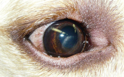
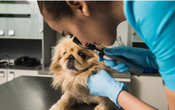
Corneal Lipidosis
What is corneal Lipidosis?
Corneal lipidosis is an accumulation of fatty substances (usually cholesterol) within the layers of the cornea.
What causes corneal lipidosis?
There are three main causes of corneal lipidosis: corneal dystrophy, corneal degeneration, and elevated blood cholesterol levels.
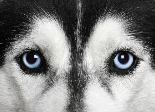
Corneal dystrophy is an inherited, or genetic condition and is most commonly seen in dogs. This condition is rarely seen in cats. It is usually present in both eyes. It is not painful and has a minimal effect on vision. Some commonly affected breeds include Beagles, Cavalier King Charles Spaniels, Siberian Huskies, Alaskan Malamutes, Samoyeds, American Cocker Spaniels, Labrador Retrievers, and Collies.
Corneal degeneration occurs secondary to inflammation in the eye and is usually associated with other eye diseases, such as anterior uveitis (inflammation of the iris, choroid, and ciliary body), keratitis (inflammation of the cornea), or scleritis (inflammation of the sclera or white of the eye) Sometimes lipid accumulation is associated with trauma, such as after corneal ulceration that healed with lipid deposits. This is seen more frequently in dogs than cats.
Lastly, elevated cholesterol levels (hyperlipidemia) can cause corneal lipidosis. This may be due to underlying causes such as Cushing’s disease, long-term steroid use, diabetes, or hypothyroidism.
What are the clinical signs of corneal lipidosis?
Lipid deposits in the cornea appear as well-defined areas of sparkly, shiny, or crystalline material. When lipidosis is due to corneal degeneration, other clinical signs may include inflammatory indicators such as eye redness or cloudiness in the eye.
How is corneal lipidosis diagnosed?
Diagnosis is based on a thorough eye examination, including fluorescein dye application, and Schirmer tear test; blood tests, age, breed, and your pet’s history. If your veterinarian suspects hyperlipidemia, then fasting cholesterol, triglyceride, and blood glucose level tests will be recommended.
How is corneal lipidosis treated?
Treatment for corneal lipidosis depends on the cause.
Corneal dystrophy does not require treatment. Your veterinarian will monitor your pet’s eyes periodically to watch for the development of corneal ulcerations.
Corneal degeneration requires treating the primary inflammatory condition in the eye which may include antibiotic or anti-inflammatory eye drops. Your veterinarian may also prescribe pain medication or artificial tear ointment to provide lubrication to the eye and comfort if the corneal surface is irregular.
Lastly, any condition that is causing an elevated cholesterol level must be treated directly in order to reduce the blood cholesterol levels. Dietary management, along with supplementation with additives to reduce cholesterol (flaxseed oil, oat bran, and niacin) can also be helpful in reducing the cholesterol levels.
“If vision is affected or if corneal ulceration occurs multiple times, referral to a veterinary ophthalmologist is recommended.”
If vision is affected or if corneal ulceration occurs multiple times, referral to a veterinary ophthalmologist is recommended.
What is the prognosis of corneal lipidosis?
Corneal dystrophy may resolve on its own and typically does not progress. It usually does not interfere with vision.
The prognosis for corneal degeneration depends on the underlying eye disease. It may progress with chronic inflammation and vision may be affected with advanced disease. Typically, corneal lipidosis does not progress after trauma.
If an underlying disease condition (Cushing’s disease, diabetes, or hypothyroidism) is identified and managed, the prognosis is good. Your veterinarian may recommend periodic blood tests to monitor cholesterol and triglyceride levels.
Contributors: Rania Gollakner, BS, DVM
© Copyright 2019 LifeLearn Inc. Used and/or modified with permission under license.

Smoke Inhalation in Dogs
What is smoke inhalation?
Smoke inhalation injuries can occur with exposure to smoke in large or small quantities. Fires produce a variety of damaging substances, each of which can affect a pet’s airways, lungs, and overall well-being.
Factors that play a role in smoke inhalation include:
• Carbon monoxide. Carbon monoxide is a colorless, odorless gas that is produced in fires. When carbon monoxide is inhaled and enters the bloodstream, it interferes with the body’s ability to deliver oxygen to organs and tissues.
• Hydrogen cyanide. This substance is released when plastic and other synthetic materials burn. Like carbon monoxide, hydrogen cyanide interferes with the body’s usage of oxygen.
• Chemical irritants. Smoke contains a number of chemical irritants. These irritants can trigger airway inflammation and constriction, as well as other lung damage. The irritants found in smoke may vary, depending on the substances that are being burned.
• Thermal injury. The heat contained within smoke can lead to burns within the airways and lungs, triggering severe swelling and inflammation.
What are the signs of smoke inhalation?
Signs of smoke inhalation vary, depending on how much smoke was inhaled, how long the dog remained in smoky conditions, what chemicals were present in the smoke, etc. A pet exposed to small amounts of smoke for a brief period of time will have very different clinical signs than a dog confined in an extremely smoky room for a prolonged period of time.
“The most common sighs of smoke inhailation are respiratory signs.”
The heat and irritants contained within smoke can also cause significant damage to the eyes. Dogs may squint due to pain, the third eyelid may remain up over the eye, and you may also notice inflammation and redness of the eyes (conjunctivitis). Burns may be seen around the face and muzzle. These burns may blister, or may appear as reddened, inflamed areas. If the nostrils are burned, you may notice nasal discharge or visible blisters within the nostrils. If the brain is deprived of oxygen due to smoke inhalation, pets may show neurologic signs. Neurologic signs are common with carbon monoxide inhalation. Signs include weakness, ataxia (impaired coordination, or walking as if drunk), seizures, and/or coma. Dogs may drool excessively or vomit. Dogs with carbon monoxide inhalation also tend to develop cherry red discoloration of the gums.
What tests will my veterinarian perform on my pet?
Your veterinarian will probably begin with pulse oximetry, bloodwork, and radiographs (X-rays) of the chest.
A pulse oximeter is a device that is used to assess your dog’s blood oxygenation. This helps to determine how efficiently your dog’s lungs are working and how effectively your dog is delivering oxygen to his organs and tissues. Pulse oximetry may be ineffective however, in cases of carbon monoxide exposure.
Bloodwork abnormalities can help your veterinarian determine the severity your pet’s lung injuries. Bloodwork findings can guide treatment decisions for your pet, while also providing information to better predict your pet’s prognosis.
Radiographs (X-rays) allow your veterinarian to determine the extent of lung damage that has occurred. In most cases, these changes are visible immediately. In many cases however, these changes will continue to progress over 48-72 hours after the initial injury. Radiographs are often repeated several times during the first 72 hours of treatment, providing a way for your veterinarian to monitor your pet’s lung damage and adjust treatment as needed.
An electrocardiogram (ECG) may also be performed, to assess your pet’s heart rate and rhythm. Oxygen deprivation can lead to heart damage, which may be detected using the ECG.
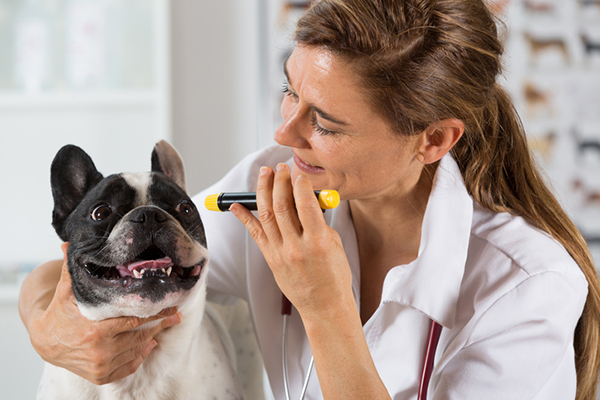
The heat associated with smoke inhalation and fires often results in ulceration of the eyes. Therefore, your veterinarian may perform a corneal stain or other ophthalmologist tests to assess your dog’s eyes and rule out injuries such as corneal ulceration.
How is smoke inhalation treated?
The treatment of smoke inhalation depends upon the severity of your dog’s signs. In most cases, the injuries seen with smoke inhalation progress over 48-72 hours; therefore, you can expect that your dog will probably be hospitalized and monitored for at least 72 hours.
“The treatment of smoke inhalation depends upon the severity of your dog’s signs.”
In the early stages, most cases of smoke inhalation are treated with oxygen therapy. Oxygen clears carbon monoxide from the bloodstream, improving oxygen delivery to the body’s tissues. Your dog may be placed in an oxygen cage, where concentrated oxygen can be delivered in a way that is non-stressful. If your veterinarian does not have an oxygen cage, they may administer oxygen via facemask or ‘flow-by’ oxygen (holding an oxygen tube near your dog’s face). If your dog has extreme upper airway swelling that prevents breathing, your veterinarian may need to place a tracheotomy tube (breathing tube inserted through the trachea) to administer oxygen.
Your veterinarian will also likely place an intravenous (IV) catheter in your dog. This catheter allows IV fluids to be administered in order to keep the lungs moist and decrease the risk of complications. Additionally, your dog may be unwilling or unable to drink water for the first few days after smoke inhalation, so IV fluids can be used to prevent dehydration.
Smoke inhalation often results in painful burns, so your veterinarian will also administer pain medication to your dog. Affected dogs are typically given injectable pain medications to keep them comfortable. Oral anesthetic rinses may also be used, if burns within the mouth are present.
“Smoke inhalation often results in painful burns, so your veterinarian will also administer pain medication to your dog.”
Your dog may also receive bronchodilators to help keep his airways open, if needed. In some cases, antibiotics may be required to treat secondary infections that can occur following lung damage.
In some cases, dogs may require additional supportive care, such as nebulization and coupage. These techniques can be used to deliver medication directly to the lungs and to remove lung secretions. See the handout “Techniques for Nebulization and Coupage in Dogs” for more information on these techniques.
What is the prognosis with smoke inhalation?
Most dogs with smoke inhalation have a good prognosis. Estimated survival rates for animals without skin burns are approximately 90%, according to multiple studies.
Dogs that present with neurologic signs, extensive skin burns, or whose condition deteriorates on the second day of hospitalization typically experience worse outcomes. The prognosis is guarded for these patients.
Contributors: Catherine Barnette, DVM
© Copyright 2019 LifeLearn Inc. Used and/or modified with permission under license.