How Can Pets Benefit From Acupuncture Treatment?

Have you ever heard of veterinary acupuncture? You may be surprised to find out that this type of treatment is available to pets and might even be a good solution for your pet’s health and well-being.

Read through the information below to find out more about veterinary acupuncture and how it can benefit pets. From there, you can choose whether to pursue this possibility for your pet. If you have any questions, feel free to call Long Island Veterinary Specialists at (516) 501-1700.

Defining Acupuncture

Acupuncture is one treatment offered in the field of Traditional Chinese Medicine (TCM). TCM is grounded in the philosophy that the body’s life force, or Qi (“CHē”), flows along body channels known as meridians. An imbalance in one’s Qi is thought to cause disharmony and, as a result, disease. Acupuncture and the action of needle insertion along specific points on the meridians stimulate beneficial effects.

Benefits of Acupuncture for Pets

Benefits of acupuncture treatment sessions may include:

  • Pain relief
  • Nerve stimulation
  • Increased blood flow to specific areas of the body
  • Muscle spasm relief
  • Release of endorphins
  • Release of cortisol

Acupuncture for Pets

Acupuncture for pets works the same way that it does for humans, including the insertion of needles. Pets can enjoy a wide variety of results when they receive acupuncture, including relaxation, relief from cramps and pain, improved mobility, reduced inflammation, and much more. Many pets respond well to this type of treatment over time. Some pets may even relax so deeply during their acupuncture treatments that they fall asleep for the duration of the treatment session.

What Acupuncture Can Treat

Acupuncture is most used to treat pain related to arthritis and other inflammatory conditions. However, it can also treat other types of pain and disease. It is often recommended as a good course of action for pets who are predisposed or who develop orthopedic conditions such as hip dysplasia.

Acupuncture can also be used to treat hot spots and benign granulomas on a dog’s legs and feet. The procedure works for these issues by relieving the nerve pain that causes dogs to lick constantly and aggravate these skin conditions.

Acupuncture may benefit pets with conditions including:

  • Chronic musculoskeletal problems – including arthritis, intervertebral disc disease, and neck and back pain
  • Post-surgical pain
  • Nerve conditions
  • Traumatic injuries
  • Slowed gastrointestinal motility, and other GI issues
  • Hip dysplasia
  • Limping
  • Seizures
  • Diarrhea and vomiting
  • Allergies
  • Itching and scratching
  • Behavioral problems
  • Heart failure and heart murmurs
  • Kidney disease
  • Liver disease
  • Cancer
dog face
dog back with needles

How a Veterinary Acupuncture Appointment Works

First, the pet will be evaluated and checked for any signs of worsening conditions or problems related to previous treatments. The pet’s health history will be recorded and any changes since the last appointment will be noted as well. From there, the problem areas to focus on will be pinpointed and the vet will talk with you about the type of treatment your pet will receive during this visit.

The process will then begin, with pets only receiving sedatives if they absolutely must have it. The needles will be inserted slowly and carefully, and your pet will be monitored throughout the experience. The process will take about 30 minutes to complete.

What to Expect Afterward

After the visit is over, chances are good and that your pet will not show any signs that anything happened at all. However, some pets may be very tired for about a day after an acupuncture appointment and will need extra rest. Other pets may be a little bit stiffer than they normally are for the first day but should have improved mobility in the days following.

Make sure your pet is allowed to rest as much as they want to after their appointment and give them plenty of cool, clean water to drink. Feed them normally afterward as well.

How to Know if Your Pet is a Good Candidate for Acupuncture

If your pet is dealing with chronic pain related to joint problems, birth defects, or arthritis, they may be a great candidate for acupuncture. Pets who have nausea, inflammation, and loss of appetite related to chemotherapy may also be ideal candidates for this treatment, as are pets who cannot receive normal pain medication due to health problems such as those with liver or kidney disease. Acupuncture may also provide pain relief and comfort for older pets who are not good candidates for orthopedic surgery.

Acupuncture is an extremely safe treatment when performed by a veterinarian who is certified in veterinary acupuncture. These treatments are a form of alternative therapy that can often be used in conjunction with your pet’s primary care to provide a holistic, comprehensive approach to certain medical conditions.

There is a lot to consider when you are trying to decide whether veterinary acupuncture is a good option for your furry friend. If you have any further questions or concerns, you can always speak with your primary veterinarian for more information or for a referral to a certified veterinary acupuncturist (CVA). For more information about Acupuncture and the Integrative Medicine services at LIVS, give us a call today at (516) 501-1700.

Spontaneous Pneumothorax


Spontaneous pneumothorax can be seen in both dogs and cats and occurs with no history of trauma or iatrogenic penetration into the thoracic cavity. The normal physiologic negative pressure within the thorax that maintains inflation of the lungs is lost, air will accumulate within the thorax and the lungs will collapse. There are two access routes allowing air into the thorax that will result in a spontaneous pneumothorax: the airways and the esophagus. The first occurs when there is 
compromise of lung or tracheal tissue resulting in air escaping into the thoracic cavity. Compromised lung tissue leading to a spontaneous pneumothorax is often caused by bullae or bleb formation at the edges of the lung lobes. These are tiny air-filled sacs that can rupture within the thorax (Figure 1). Bullae and blebs are most frequently found in dogs with no concurrent lung disease and it is the most common cause of spontaneous pneumothorax. Bullae and blebs are less common in cats, but they are the main cause of feline cases of spontaneous pneumothorax.

The second route of entry into the thorax is from esophageal perforation that results in both pneumothorax and additional air accumulation in secondary locations causing subcutaneous emphysema or pneumomediastinum. Secondary causes of spontaneous pneumothorax in dogs include cancer, bacterial pneumonia, fungal infection, pulmonary abscesses, grass awn migration, pulmonary thromboembolism, and heartworm disease. In cats, spontaneous pneumothorax can be caused by inflammatory airway disease, heartworm or lungworm infection, and bronchopulmonary dysplasia. In some patients the cause is unknown.

Spontaneous pneumothorax occurs primarily in deep-chested, large breed dogs with no sex predilection. Siberian Huskies and other Northern breeds are overrepresented. There are no known common associations in cats. Clinical signs noted in dogs include increased respiratory rate, cough, anxiety, cyanosis, overinflation of the thorax, and an orthopneic posture. Common signs noted in cats are respiratory distress, cough, collapse, lethargy, anorexia, and vomiting.

Initially, we recommend 3-view thoracic radiographs to assess for pneumothorax (Figure 2). This diagnostic test rarely confirms the underlying cause and additional advanced imaging such as a CT scan often becomes necessary (Figure 3). Before referral, a therapeutic thoracocentesis along the dorsolateral thorax is necessary to stabilize the patient, as well as supplemental oxygen therapy. Once the patient arrives at our facility, we will reassess with thoracic radiographs or TFAST for an additional accumulation of air. Before the CT scan, we will do an additional thoracocentesis or place chest tubes to keep air out of the cavity and allow the lungs to fully expand for imaging. Supplemental oxygen will be necessary at all times to assist with increased oxygen delivery to the body.

Treatment options vary depending on the severity of the clinical signs. The underlying cause of spontaneous pneumothorax dictates which treatment option will be most appropriate. Most often, spontaneous pneumothorax cannot be managed medically (oxygen supplementation and chest tube management) long-term, and surgical intervention is warranted to remove the primary cause of the pneumothorax.

Surgical treatment most often includes a lung lobectomy. This can be accomplished using three different approaches to the thorax: minimally invasive thoracoscopy, median sternotomy, or lateral thoracotomy. The choice of the approach depends on the underlying cause of the disease and the location of the affected lung lobe. If laterality is determined via the CT scan then a thoracotomy is the best option. If we cannot determine the cause then a median sternotomy is chosen. Once the thoracic cavity is open, each lung lobe is inspected for an abnormality causing a leak. If no overt abnormality is noted, the thoracic cavity is filled with warm saline; bubbles will appear in the saline from the lung lobe that is affected. Stapling equipment is used to remove the lung lobe at the base. The lobe will be submitted for histopathology. The warm saline will be used again to assess the remaining bronchus for any leaks. If a chest tube was not placed before surgery, one is placed before closing the thorax.

We will monitor chest tube production in the ICU postoperatively. Once air and fluid are no longer accumulating (and being removed via the tube), the tube is removed and the patient will most likely be discharged the next day.

Long-term outcomes for dogs with spontaneous pneumothorax are excellent with surgical intervention and lung lobectomy. Very rarely are cases not amenable to surgical resection and those patients’ prognoses vary depending on the severity of the disease. Recurrence rates of spontaneous pneumothorax are approximately 3% with surgical treatment and as high as 50% with medical treatment. Mortality rates reflect the need for appropriate surgical management. The mortality rate with surgery is 12% and is over 50% with medical management.

Outcome data for cats is more limited. The prognosis can still be good overall depending on the underlying cause and response to therapy. In a previous study, 54% of the cats evaluated survived to discharge with the majority being treated medically. For cats with small volume pneumothoraces, conservative therapy with oxygen and chest tube /thoracocentesis may offer a good outcome. Surgery may be needed in severe cases or when there is persistent pneumothorax present despite medical management.

Figure 1 – A bleb is noted along the periphery of the lung lobe.


Figure 2 – Left lateral radiograph of a dog with a pneumothorax. Note the collapse of the lung lobe (thin arrow) and the raised heart.

Figure 3 – A sagittal view of a CT scan of a dog with a bleb on the periphery of the lung lobe (circle).

Article Written By:
Catherine Loughin, DVM
DACVS, DACCT
Soft Tissue, Oncologic, Orthopedic, Neurosurgery

Cranial Cruciate Ligament Repair: Tibial Plateau Leveling Osteotomy (TPLO)

One of the most common injuries to the knee of dogs is tearing of the cranial cruciate ligament (CCL). This ligament is similar to the anterior cruciate ligament (ACL) in humans. There are actually two cruciate ligaments inside the knee: the cranial cruciate ligament and caudal cruciate ligament. They are called cruciate because they cross over each other inside the middle of the knee.

When the CCL is torn or injured, the shin bone (tibia) shin bone slides forward with respect to the thigh bone (femur), which is known as a positive drawer sign. Most dogs with this injury cannot walk normally and experience pain. The resulting instability damages the cartilage and surrounding bones and leads to osteoarthritis (OA).

Call Us

What options are there for repairing my dog’s torn CCL?

When the cranial cruciate ligament is torn, surgical stabilization of the knee joint is often required, especially in larger or more active dogs. Surgery is generally recommended as quickly as possible to reduce permanent, irreversible joint damage and relieve pain.

“Surgery is generally recommended
as quickly as possible to reduce
permanent, irreversible joint damage
and relieve pain.”

Several surgical techniques are currently used to correct CCL rupture. Each procedure has unique advantages and potential drawbacks. Your veterinarian will guide you through the decision-making process and advise you on the best surgical option for your pet.

My veterinarian is recommending a tibial plateau leveling osteotomy to fix my dog’s torn CCL. What does this surgery involve?

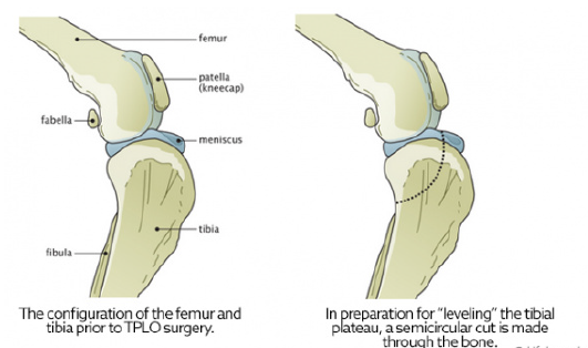
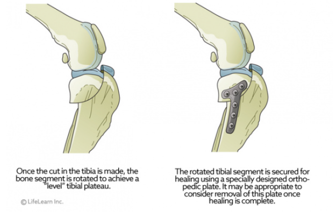
A major advancement in the treatment of CCL rupture has been the development of tibial plateau leveling osteotomy or TPLO. This surgery changes the angle and relationship of the femur and the tibia. The overall intent of the surgery is to reduce the amount that the tibia shifts forward during a stride. This is accomplished by making a semicircular cut through the top of the tibia, rotating the top of the tibia, and using a bone plate to allow the tibia to heal. This realignment of the surfaces within the stifle (knee) helps to provide stability during a stride and helps to reduce future joint inflammation and OA. By carefully adjusting the angle or slope of the top of the tibia, surgeons are able to replicate a more normal configuration of the knee joint and reduce mechanical stress.

“A major advancement in the treatment of CCL injuries has been the development of TPLO.”

To better understand the purpose of leveling the tibia, we often use the analogy of a wagon on a hill. Imagine a wagon tied to a post on the slope of a hill. As long as the rope holds, the wagon does not roll downhill. If we add excess weight (or downward force) to the wagon, the rope could break, and the wagon will roll down the hill. However, if the wagon is on level ground, it will not roll forward with added weight, even if the rope is broken.

In this example, the wagon is the femur, and it slides down slope of the top of the tibia when the CCL is ruptured. This instability leads to damage and destruction of the cartilage and bones of the knee joint. TPLO surgery levels the tibia to prevent the femur from sliding forward, thereby stabilizing the
joint.

TPLO surgery involves making a curved cut in the tibia from the front to the back, much like half a smiley face. The top section of the tibia is then rotated backward until the angle between the tibia and femur is deemed
appropriately level, typically between 2 and 14 degrees, with 5 degrees being the ideal angle. A metal bone plate is then used to affix the two sections of tibia in the desired positions, allowing the tibia to heal in its new configuration.

How long will it take for my dog to recover from TPLO surgery?

Healing from TPLO surgery is generally rapid.

  • About half of all canine patients will begin walking on the injured leg within 24 hours after surgery.
  • At 2 weeks postoperatively, most dogs are bearing moderate to complete amounts of weight on the affected leg.
  • By 10 weeks, most dogs do not have an appreciable limp or gait abnormality.
  • As mentioned above, at 4 months postoperatively, the majority of dogs can begin walking and playing normally, with only the stressful activities restricted.
  • Within 6 months, most dogs can resume full physical activity.

Pain management during and after stifle (knee) surgery is critical, so be sure to give all medications as prescribed and use them until they are gone. Physical rehabilitation post-operatively will speed healing. Ask your veterinarian about incorporating rehabilitation into your dog’s recovery plan.

“Pain management during and after stifle (knee) surgery is critical, so be sure to give all medications as prescribed and use them until they are gone.”

The most common complication after TPLO is infection. Studies conclude that infection occurs in less than 10% of all patients, with many surgeons reporting much lower complication rates. Your dog will need several recheck examinations and radiographs (X-rays) to ensure that the area is healing properly.

Contributors: Tammy Hunter, DVM; Robin Downing, DVM, DAAPM, DACVSMR, CVPP, CRPP
© Copyright 2019 LifeLearn Inc. Used and/or modified with permission under license.